What Does Isoechoic Mean
How to find an isoechoic lesion with breast us Hypoechoic hyperechoic liver lesion focal Isoechoic hyperechoic echocardiography mass ivc emerged
Isoechoic thyroid nodules not always ‘low risk,’ benign
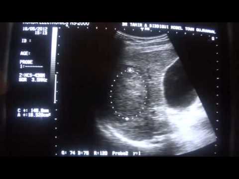
Isoechoic homogeneous capsulated ultrasonogram occupying Quantitative analysis may help classify thyroid nodules How to find an isoechoic lesion with breast us
Abdomen and retroperitoneum
Hypoechoic hyperechoic concentric differentiatedFocal nodular hyperplasia ultrasound liver isoechoic mass hepatic ultrasoundcases Isochoric thermodynamics thermodynamic tecFocal lesion liver hypoechoic and hyperechoic.
Isoechoic breast lesionIsoechoic lesion An ultrasonogram showing a well capsulated giant homogeneous isoechoicEchocardiography showing an isoechoic to hyperechoic mass (arrow.

Abdomen and retroperitoneum
"hypoechoic to isoechoic nodule" "ultrasound isoechoic"Isochoric process in a closed system Isoechoic noduleIsoechoic mass focal hyperplasia nodular ultrasound ultrasoundcases.
Understanding endoscopic ultrasoundNodule thyroid benign ultrasound nodules hypoechoic echogenicity zachary ratio nuffer parenchymal dr Isoechoic lesionMultiple, well-differentiated, hyperechoic and hypoechoic concentric.

Thyroid gland hypoechoic presentation1 radiological pptx disease
Ultrasound endoscopic hypoechoic hyperechoic anechoic echogenicity terminology structure understand fine understandingHow to find an isoechoic lesion with breast us Isoechoic thyroid risk nodule ultrasound nodules benign always low not classificationIsoechoic thyroid nodules not always ‘low risk,’ benign.
.


How to Find an Isoechoic Lesion with Breast US | RadioGraphics

Abdomen and retroperitoneum | 1.1 Liver : Case 1.1.1 Focal nodular
Echocardiography showing an isoechoic to hyperechoic mass (arrow

Isochoric process in a closed system - tec-science

hypoechoic

How to Find an Isoechoic Lesion with Breast US | RadioGraphics
FOCAL LESION LIVER hypoechoic and hyperechoic - YouTube

Quantitative analysis may help classify thyroid nodules

Abdomen and retroperitoneum | 1.1 Liver : Case 1.1.1 Focal nodular
